Nuclear magnetic resonance (NMR) is a cornerstone technique in chemistry, physics, and biology, pivotal for non‑invasive study of the structure and dynamics of molecules within the human body. Among its primary applications is magnetic resonance imaging (MRI), an indispensable tool in modern medicine that allows doctors to easily observe pathological changes in patients. Our research team in the Institute of Microelectronics (IME) at the University of Macau (UM) has recently developed a compact MRI scanner based on complementary metal‑oxide‑semiconductor (CMOS) technology, aiming to advance personalised medicine.
The advent of multi-nuclei MRI technology
With the advent of multi-nuclei MRI technology, we can now perform medical imaging using various nuclei, such as hydrogen-1 (1H) and fluorine-19 (19F), to improve the effectiveness of diagnosis and treatment monitoring. Hydrogen-1, which is abundant in biological tissues, allows for clear anatomical MRI images. However, its ubiquity limits its effectiveness in tracking specific substances within the body. In contrast, fluorine-19 is extremely rare in biological tissues, making it an excellent marker for tracking targeted drugs and other specific substances with high precision, thereby allowing a more accurate assessment of disease progression and treatment efficacy.
Innovations in the miniaturisation of MRI technology
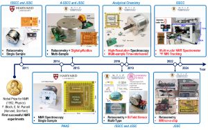
Traditional MRI machines, which use superconducting magnets and discrete electronic components, are bulky, expensive, and have limited applications. However, the advent of miniaturised NMR/MRI systems is changing the landscape. These new systems are powered by CMOS application-specific integrated circuits (ASICs) and more affordable permanent magnets. In the initial stages, our research team focused on miniaturising NMR spectroscopy and relaxometry, known as NMR-on-a-chip. In 2023, we developed the first three-dimensional MRI-on-a-chip platform, and introduced the miniaturised MRI scanner.
Creating a miniaturised multi-nuclei NMR/MRI platform
Over the past decade, the research team in the State Key Laboratory of Analog and Mixed-Signal VLSI at UM has been working on the miniaturisation of NMR/MRI systems and has developed several generations of portable NMR/MRI platforms, each with unique and innovative features. Among these platforms, the latest generation is the first miniaturised multi-nuclei NMR/MRI platform with a customised silicon chip for ex vivo 19F MRI tracking.
The scanner on this platform is equipped with a 0.52 Tesla permanent magnet and a highly integrated high-voltage insulated silicon ASIC. It is designed to be compact and lightweight, with optimised size, weight, imaging area, image resolution, and signal-to-noise ratio.
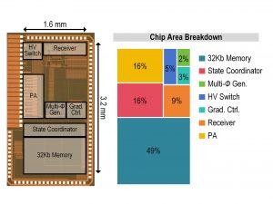
At the core of this miniaturised NMR/MRI system is a customised CMOS ASIC chip. The compact core area is only 4.1mm², which enhances the portability and sensitivity of the miniaturised multi-nuclei NMR/MRI platform. Its components include: 1) an arbitrary pulse sequence synthesiser (equipped with 32kb of memory and a state coordinator to instruct the operation of each module); 2) a high-voltage transmitter (TX, which sends RF signals to the coil to excite the nuclei in the sample); 3) a low-noise receiver (RX, which captures and processes signals from the nuclei); 4) a pair of high-voltage-tolerant switches (which protect RX from TX); and 5) a three-dimensional gradient controller (comprising three sets of 12-bit digital-to-analogue converters used to programme the necessary gradients for spatial encoding of the nuclei).
Pushing technological boundaries
The miniaturised multi-nuclei NMR/MRI platform employs composite radio-frequency (RF) pulses. These pulses are a combination of RF signals with precisely adjusted phases and amplitudes, designed to minimise off-resonance effects, which is critical for multi-nuclei NMR/MRI. In addition, these pulses can mitigate the effects of inhomogeneous static and RF magnetic field strengths, such as image quality degradation. It is worth noting that such composite pulses require a phase resolution of less than 1°, which most compact NMR/MRI systems are unable to achieve. To address this limitation, we have integrated a multi-phase generator based on a phase-interpolated dual-DLL architecture and improved the accuracy of the NMR spectrum amplitude by 28 per cent, significantly enhancing the quality and capabilities of our low-field NMR platform.
This transformative technology not only revolutionises the generation of composite RF pulses but also improves the power transmission mechanism within the system. By leveraging a class-D power amplifier equipped with a sophisticated high-voltage NMOS array, the system can deliver the high-power capacity required for large FOV MRI applications (20.5W into a 100-Ω load). The high-voltage-tolerant switches also play a crucial role during nuclear excitation as they protect the receiver and reduce noise during acquisition, thereby ensuring optimal signal quality and system performance.
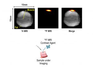
In the medical field, 19F tracking is a hallmark application of multi-nuclei MRI. To evaluate the performance of this new system, we used perfluorocarbon (PFC) as an MRI contrast agent and injected it into the ex vivo porcine sample to obtain 1H/19F images. The 1H image details the structure of the porcine sample (muscle and fat), while the 19F image shows the location of the PFC. We found that combining these images allows precise tracking and analysis of specific chemical substances within the sample. The application of this technology in a miniaturised multi-nuclei NMR/MRI system marks a milestone in our research and development efforts.
Advancing personalised medicine
We believe that this compact, miniaturised multi-nuclei NMR/MRI system will drive advancements in personalised medicine. By enabling in-depth molecular analysis of body tissues, the system has the potential to revolutionise early disease detection, treatment strategies, and disease management. As our team continues to make strides in CMOS integrated circuit technology, we are setting the stage for a future where this sophisticated imaging technology becomes a standard tool in healthcare, offering medical professionals more powerful diagnostic capabilities than ever before.
Authors:
Fan Shuhao obtained her PhD from the University of Macau in 2024. She earned her BSc degree from Northeast Forestry University in 2016, and her MSc degree from the Hong Kong University of Science and Technology in 2017. She was also a visiting student at Harvard University in 2024. Her research interests include portable NMR/MRI platforms with customised integrated circuits, and her work has been featured in conferences and journals such as the IEEE International Solid-State Circuits Conference, IEEE Journal of Solid-State Circuits, IEEE Transactions on Circuits and Systems I: Regular Papers, and the International Conference on Miniaturized Systems for Chemistry and Life Sciences.
Lei Ka Meng has been an assistant professor in the Institute of Microelectronics at the University of Macau (UM) since 2019. He received his BSc and PhD degrees from UM in 2012 and 2016, respectively. His research interests include precision analogue integrated circuits, sensors and analogue front-end interfaces, and high-resolution portable NMR/MRI platforms.
Text & Photo / Fan Shuhao, Lei Ka Meng
Chinese Translation / Davis Ip
Source: UMagazine ISSUE 30

The miniaturisation of NMR/MRI systems over the past decade

The customised integrated circuit for the miniaturised multi-nuclei NMR/MRI platform

MRI tracking of 1H and 19F in the porcine sample injected with PFC, which helps differentiate between muscle (T1: 191.2 ms) and fat (T1: 82.9 ms) in the sample
